Vision
- Details
- Published: Monday, 02 April 2012 17:25
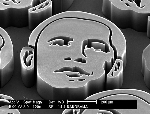
Microscopic faces of Barack Obama made using nanotechnology, and imaged using a scanning electron microscope. Each face consists of millions of vertically-aligned carbon nanotubes, grown by a high temperature chemical reaction. (Image source: NANOBAMA_face1; www.nanobama.com by John Hart, 2013)
Within the past decades, advances in miniaturization from micro to nano-scale have had dramatic impacts on our lives. Consumer electronics, which once occupied large volumes, now fit in the palm of a hand. But nanotechnology does not only improve electronics. Also material sciences, chemical engineering or biology are strongly profiting from nanotechnology. The tremendous achievements in all of these areas would not have been possible without corresponding material analytics techniques.
To date it is not possible to get a comprehensive representation of a specimen including internal and external 3D-structure analysis as well as a chemical analysis without destroying the sample. In this respect nano-scale material analytics is currently on the edge of a new era, which is targeted in NanoXCT. The project addresses the limitations of conventional techniques using 3D X-ray computed tomography, which allows for a non-destructive and fully three-dimensional characterization of specimens. In order to facilitate X-ray computed tomography at the nanometer scale, NanoXCT comprises a novel concept of an ultra-bright X-ray source in combination with a high precision focusing and emission system. Furthermore, a highly sensitive, wide field-of-view and small pitch X-ray detector concept will be included. The concept is perfected by a high precision manipulation system, which allows for alternative scanning geometries such as helical CT and a suitable software environment for data processing and analysis. The system is completed by the X-ray fluorescence elemental composition analyzing unit.
| Specimen diameter (mm) | Field of view (mm) | Voxel size (nm) |
| 0.2 | 0.175 | 50 |
| : | : | : |
| 1.0 | 1.0 | 285 |
Table 1: Targeted geometric specifications of the NanoXCT system.
NanoXCT links the activities of 10 partners from 7 European countries including 5 SMEs to design, develop and implement a desktop X-ray computed tomography system for non-destructive characterization of nano materials and components. The targeted specifications yield a wide field-of-view of up to 1 mm, a specimen size of up to 1 mm and a voxel size down to 50 nm.